Introduction
How is Flow Cytometry Performed?
The Hull of the Flow Cytometer Apparatus
Flow Cytometry in the World of Hematology
Flow Cytometry in Leukocyte Analysis
References
Cell processes are complex and are controlled through dynamic intracellular signaling. To better understand these processes, flow cytometry is used to phenotype cells by taking their dimensions and assaying their interiors in a streamlined fashion.
Flow cytometry is a technique used in Hematology (the study of blood) to sort cells in accordance with their size and complexity. Since its conception in 1976, flow cytometry has acquired new functions. It can now sort cells by fluorescence-based approaches to analyze a cell's viability and more.

Image Credit: Buravleva stock/Shutterstock.com
How is Flow Cytometry Performed?
First, the cells that are being targeted are fixed using formaldehyde, immobilizing proteins, and preserving transient signals. Then, the fixed cells are made permeable through a detergent, allowing the passage of antibodies between the intercellular space. To ensure successful antigen detection, a specified formaldehyde and detergent concentration is critical, dependent on the target epitopes' nature. After this is accomplished, the quantification of blood cells can be determined through the flowthrough of the sample and sheath fluid. This technique allows us to classify cells based upon their size and complexity and could even go as far as elucidating the stage/differentiation of fluctuating cells.
Another facet of flow cytometry presents itself as fluorescence-activated cell sorting (FACs). This specialized flow cytometry variant was brought to us in the 1960s. This technique allows the user to sort many biological cells throughout a heterogeneous mixture, all done through the light scatter of fluorescence. This is done one cell at a time and functions well because each cell has its own light scattering characteristics.
The Hull of the Flow Cytometer Apparatus
Firstly, this fluidic system uses laser modules to illuminate a sample, emitting light at the visible region (380-700nm). The cell medium introduced to the device passes through the laser, dubbed the "integration point". From here, light is scattered based on the refractive properties of anything within the cytoplasm. The principle this apparatus is based on is called hydrodynamic focusing.
Here, the sheath fluid and the core fluid flow in a streamlined motion. The sheath fluid runs at a laminar flow while the core fluid flows at a slightly higher pressure. This allows the target cells to streamline in a single file in the direction of the laser, allowing the device to focus on the core fluid.
Photomultiplier tubes are often the detector of choice in flow cytometry, which then read the light scatter emitted from the integration point. The two modes of light analyzed are forward scatter and side scatter, the patterns of which pertain to larger and smaller cells respectfully. This scattered light is converted into a voltage pulse, which embodies an analog signal that can be read.
Once these readings have been taken, the analog signal is converted to a digital one through an ADC unit, and the final digital readout can then be read on a monitor. The resulting readout takes the form of a histogram, with cell count on the y-axis and forward scatter signal on the x-axis.
Cells with higher complexity, or granularity, will have a propensity to scatter more light at more varied angles. This implies that more complex cells will lead to more side scatter, while simpler cells emit more forward scatter.
Flow Cytometry in the World of Hematology
Over the decades, flow cytometry has been used in various applications, expounding upon fields of epidemiology, genetics, and more.
If cells are thought to be abnormal or aneuploidy, flow cytometry can be used to confirm DNA content. When using propidium iodide as a fluorescent dye, it can intercalate into the helical structure of the DNA. The resulting fluorescent signal will be proportional to the content of DNA within the nucleus, allowing scientists to quantify the gain or loss of DNA empirically.
To emphasize the efficacy of this methodology, the excessive amount of DNA found in tumor cells can be determined, given that aneuploidy DNA is often linked with malignant tumors. Under this postulation, one could not only diagnose malignant cancers but identify where the highest concentration of aneuploidy cells are, effectively pinpointing tumor formation.
Flow cytometry can also be used in other fields to detect and quantify the amount of fetal red cells in maternal blood. While this could be used to diagnose pregnancy, this can also be used in predictive medicine to prevent fetal-maternal hemorrhaging (FMH). This is when fetal red blood cells invade maternal circulation, resulting in obstetric/trauma-related complications during pregnancy.
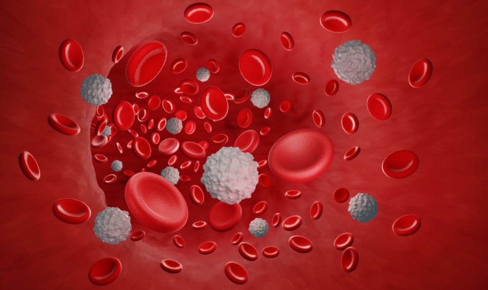
Image Credit: ART-ur/Shutterstock.com
Flow Cytometry in Leukocyte Analysis
Hundreds of thousands of patients are diagnosed with leukemia and lymphoma each year. To bypass the radiation that accompanies CT scans, immunophenotyping of leukemias/lymphomas using flow cytometry is being undergone. This can be accomplished by categorizing the varying cell surface markers, like CD markers, found amongst all hematolymphoid neoplasms.
Delving further, one can systematically discern the nature of leukemia based on the CD markers identified by the flow cytometer. For example, if the markers CD19/20/22 are positive, we know that the cancer is associated with the B cells of the immune system. If CD5 and CD23 are positive, the diagnosis is Hodgkin's lymphoma. If CD23 is negative and CD22 is positive, the diagnosis is mantle cell lymphoma, and so on. Though the instrument was assembled decades prior, modern science has found a way to implement the flow cytometers modus operandi to varying forms of analysis.
References:
- McKinnon K. M. (2018). Flow Cytometry: An Overview. Current protocols in immunology, 120, 5.1.1–5.1.11
- Picot, J., Guerin, C. L., Le Van Kim, C., & Boulanger, C. M. (2012). Flow cytometry: retrospective, fundamentals, and recent instrumentation. Cytotechnology, 64(2), 109–130
- Muñoz-García N, Lima M, Villamor N, Morán-Plata FJ, Barrena S, Mateos S, Caldas C, Balanzategui A, Alcoceba M, Domínguez A, Gómez F, Langerak AW, van Dongen JJM, Orfao A, Almeida J. (2021) Anti-TRBC1 Antibody-Based Flow Cytometric Detection of T-Cell Clonality: Standardization of Sample Preparation and Diagnostic Implementation. Cancers.; 13(17):4379.
- Hassett J, Parker J. (1995) Laboratory practices in reporting flow cytometry phenotyping results for leukemia/lymphoma specimens: results of a survey. Cytometry; 22(4):264-81; discussion 330.
- Ross JS. (1996) DNA ploidy and cell cycle analysis in cancer diagnosis and prognosis. Oncology (Williston Park);10(6):867-82, 887; discussion 887-90. PMID: 8823802.
- Gujral S, Subramanian PG, Patkar N, Badrinath Y, Kumar A, Tembhare P, Vazifdar A, Khodaiji S, Madkaikar M, Ghosh K, Yargop M, Dasgupta A. (2008) Report of proceedings of the national meeting on "Guidelines for Immunophenotyping of Hematolymphoid Neoplasms by Flow Cytometry". Indian J Pathol Microbiol; 51(2):161-6
- Julia Gala de Pablo, Matthew Lindley, Kotaro Hiramatsu, and Keisuke Goda. (2021) High-Throughput Raman Flow Cytometry and Beyond Accounts of Chemical Research 54 (9), 2132-2143
Further Reading
- All Flow Cytometry Content
- Using Flow Cytometry in Disease Diagnosis
- Flow Cytometry and Drug Discovery
- Using Flow Cytometry in Biomarker Detection
- Flow Cytometry Methodology, Uses, and Data Analysis
Last Updated: Jul 15, 2022

Written by
Vasco Medeiros
Obtaining an International Baccalaureate Degree at Oeiras International School, with higher levels in Chemistry, Biology, and Portuguese, Vasco Medeiros has just graduated from the University of Providence College with a Bachelor of Science. Before his work as an undergraduate, he first began his vocational training at the HIKMA Pharmaceuticals PLC plant in Ribeiro Novo. Here he worked as a validation specialist, tasked with monitoring the gauging and pressure equipment of the plant, as well as the inspection of weights and products.
Source: Read Full Article