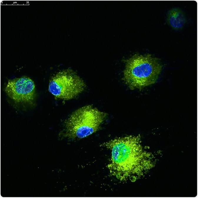
Bimolecular fluorescence complementation (BiFC) assay is a nonintrusive fluorescence-based method used to exactly visualize the in vivo interaction of proteins with the help of fixed cells or live-cell imaging. This technique is also used to localize the subcellular interacting proteins without any additional external agents.

Credit: Vshivkova/Shutterstock.com
BiFC technique is popular because of its less complexity, ease of use, and the potentiality to accomplish with standard confocal laser scanning or epifluorescence microscopy. Although this approach enjoys many advantages, there are several limitations that are needed to be considered.
Shortcomings of BiFC
Slow maturation
Numerous features of BiFC assay restrict its pertinence and changes should be done accordingly to the outcome of the assay. The most important limitation of this assay is slow fluorophore maturation, which is reflected in the chemical reaction essential for cyclic fluorophore formation. This in turn precludes detection of modifications in protein interactions by BiFC. In addition, the slow maturation process affects the visualization of short-lived or active interactions in cells.
Irreversible BiFC formation
Dynamics of interacting protein sex change and complex separation are likely to get affected by the formation of bimolecular fluorescent complex, which cannot be reversed under several in vivo conditions. The bimolecular fluorescent complex formation can, however, become reversible if rapid alterations in the fluorescence signal occur with possible degradation of proteins affecting the signals in the cells. However, while this drawback is beneficial to detect weak and transient interactions, it cannot be used to analyze kinetics of the dynamic complex.
The signal obtained from fluorescence complementation has been reduced under conditions and are expected to decrease the formation of complex in some in vitro conditions.
Additionally, fragments of fluorescent protein have limited ability to correlate with each other independent of an interaction among proteins. This main source of background signal in BiFC differs based on the characteristics of fusion protein and their expression level.
The amount of fluorescence radiating from resulting BiFC complexes is not consistent with the kinetics of protein interactions. At first, the time involved in association between fluorescent protein fragments and the chemical reactions required to mature the fluorophore leads to a delay between the start of interaction and commencement of fluorescence emission. The duration of the delay possibly depends on the fluorophore formation kinetics, and hence, when using rapidly maturing fluorescent proteins the BiFC complex fluorescence can be detected sooner.
Unsuitable for anaerobic organisms
The other limitation of the BiFC approach is that it is not suitable for anaerobic organisms, since the formation of fluorophore requires oxygen. The protein should adapt the fluorescent protein fragments without interrupting their functions or harmful effects for the cell.
Protein folding and functionality
There are a few other potential complications and unfavorable factors that should be considered for assay of BiFC. For applying BiFC assays, fluorescent proteins that are fragmented should not be able to fold together instinctively except when they are at close proximity by the interaction between two proteins.
Another significant complication of BiFC includes the viability of the fusion proteins as in other techniques that use labeled or fusion proteins. Furthermore, the intensity of restructured fluorescence fragments should be similar to that of the whole fluorescent protein and with adequate brightness to be differentiated from other background signals. However, deceptive positive fluorescent signals can also be detected.
For confirming a positive interaction between proteins, it is important to have appropriate control, for example, a binding protein should transmit mutation in the binding interface. It is also difficult to find a suitable negative control while examining the interaction between two new proteins whose structural or biochemical information is not known.
Lack of stability
Another limitation of BiFC is the lack of interaction stability it exhibits i.e., the association between the fragments of fluorescent proteins is not stable under certain conditions. Sound insight of complex dynamics of BiFC formation in live cells and enhancement of complementing fragments that cause minimal damage to the interacting protein partners might be valuable contributions.
Independent association
The inherent capacity of fluorescent fragments to correlate without depending on the interactions between the proteins that are fused to fragment is also a drawback of the technique. Based on the expression level and the protein fusion, background signal in BiFC assay differs. Enhancing fluorescent proteins to reduce inherent association tendency without lowering their capacity to interact together when brought close to each other would improve the assay less sensitive to protein expression levels.
Dependence on temperature and protein expression
One major limitation in BiFC methods is the use of lower temperatures, for example at 4°C or 25°C, at which this approach is performed well. But this is not applicable to all type of cells, as the reduced temperature might be harmful to them.
Another drawback of BiFC is its dependence on the translational fusion protein expression. The level of expression of fluorophore-tagged proteins hinders their function and orientation of the fluorophores. Hence, fusions along N-terminal or C-terminal must be taken into account in the BiFC assay.
Sources
- https://www.ncbi.nlm.nih.gov/pmc/articles/PMC4932868/
- https://www.ncbi.nlm.nih.gov/pmc/articles/PMC3169261/
- https://www.ncbi.nlm.nih.gov/pmc/articles/PMC2518326/
- https://www.ncbi.nlm.nih.gov/pmc/articles/PMC4417415/
- https://www.ncbi.nlm.nih.gov/pmc/articles/PMC2151733/
- journals.plos.org/plosone/article?id=10.1371/journal.pone.0100589
- https://www.botanik.kit.edu/botzell/downloads/Pub_Stolpe_2005.pdf
- http://pubmedcentralcanada.ca/pmcc/articles/PMC2829325/
- www.sciencedirect.com/…/bimolecular-fluorescence-complementation
Further Reading
- All Fluorescence Content
- What is Fluorescence Spectroscopy?
- How do Epifluorescence Microscopes Work?
- GFP-tagging in Fluorescence Microscopy
- Fluorescence Quenching
Last Updated: Feb 26, 2019
Source: Read Full Article


