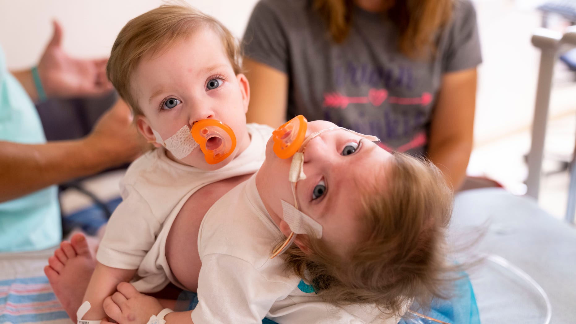
Sarabeth and Amelia were born connected from the chest down to the belly button.
(Joe Hallisy, Michigan Medicine)
A pair of formerly conjoined twins have spent the last several weeks getting to know their new normal after surgeons in Michigan successfully separated the pair in an 11-hour operation. Sarabeth and Amelia Irwin were born connected from the chest to the belly button and shared one liver, but each had their own arms, legs, hearts and digestive tracts.
Phil and Alyson Irwin, of Petersburg, Mich., said they didn’t know something was amiss with their twins until the 20-week ultrasound, which they assumed was to discover the gender of their older daughter’s new siblings.
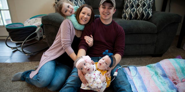
The girls, pictured at home with their parents and older sister, were born via caesarian section at 34 weeks gestation.
(Joe Hallisy, Michigan Medicine)
However, the pair told Michigan Health News, the ultrasound showed the twins were conjoined, and the obstetrician explained that they couldn’t help them and that “we had an unfortunate decision to make.”
The Irwins landed in the hands of Michigan Medicine’s Fetal Diagnosis and Treatment Center, where Dr. Marjorie Treadwell, the center’s director, met with them.
“We had some good appointments and some where we were left crying,” Alyson Irwin said, according to the blog. “We were preparing for the worst possibility.”
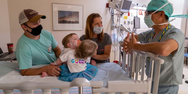
The girls’ surgical team was made up of over two dozen doctors, nurses and specialists.
(Joe Hallisy, Michigan Medicine)
At 34 weeks, the girls were delivered via caesarian section and placed on their mother’s chest before being taken to Mott Newborn Intensive Care Unit for 85 days. For the next 13 months, the girls underwent physical and occupational therapy at C. S. Mott Children's Hospital, while the separation surgery was scheduled and a plan was formulated.
OVER HALF OF CORONAVIRUS-INFECTED PREGNANT WOMEN SHOWED NO SYMPTOMS, CDC REPORT SAYS
More than two dozen doctors, nurses and specialists were on hand for the surgery, which is believed to be the first in the state, according to Michigan Health News.
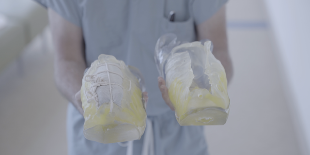
The team used 3-D printed models to help plan for separating their abdomen and their shared liver.
(Joe Hallisy, Michigan Medicine)
“For everyone in the room, it was a very emotional and extraordinary moment when the last incision was made to separate these girls from one to two,” George Mychaliska, M.D., pediatric and fetal surgery at Mott, said. “This was a monumental team effort that involved virtually every clinical department here and a group that was incredibly committed to collaborating in the most innovative way.”
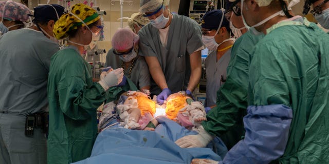
The teams color-coded their surgical caps to help determine who was working on each twin.
(Joe Hallisy, Michigan Medicine)
“We all recognized that in some way, psychologically, this might be traumatic for these little patients because their lived experience had been with their twin right next to them,” Mychaliska told the health blog. “Our pediatric intensive care unit configured a room that could support both of them so they would see each other when they woke up.”
Once the surgery was deemed a success, the girls’ parents began imagining a series of firsts they would get to experience with their separated twins, like rocking each girl to sleep separately and watching them crawl and walk on their own.
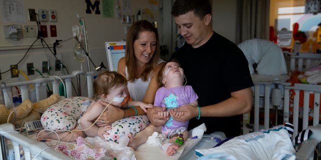
The girls spent several weeks in the intensive care unit recovering from the surgery.
(Joe Hallisy, Michigan Medicine)
“We always knew that we had to give them the chance to live their lives independently but it was so hard to imagine them going through such a big surgery,” Alyson Irwin told the health blog.
Source: Read Full Article


