By Jeyashree Sundaram, MBA
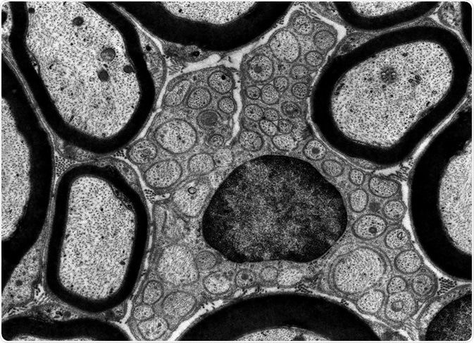
A significant technology, Transmission electron microscopy (TEM) has found its way into cell biology since 1940. When compared with light microscopes, TEM can achieve a very high resolution due to the shorter wavelength of the electron beam it uses. This feature enables TEM to visualize the structure of the cells such as membrane systems, cilia, organelles and its complexity. Regardless of the advantages, TEM has its own limitations.

Credit: Jose Luis Calvo/ Shutterstock.com
Sampling abilities
Any high-resolution imaging technique has its own in-built limitation: at any given time one can examine just a small part of a specimen; the higher the resolution, the lower will be its sampling capabilities.
All TEMs may have been used to analyze no more than of sample material till date. A researcher must get to know the basic aspects of one’s specimen before attempting to know the finer aspects.
Projection limitations
TEM works in such a way that we see 2D images of 3D specimens, viewed as part of transmission. Because our eyes and brain understand reflected light routinely we may assume that we may be able to interpret images of TEM, but this is not true.
For example, when we see a picture of two animals facing away from each other, it may appear (two-dimensionally) as a single body with two heads at either end but we still understand the picture, as we understand the true nature of the animals in a 3D scenario. in contrast, when we see a similar misleading TEM image, we may not interpret it correctly, and there may also be artifacts in TEM images.
This specific drawback in TEM is termed as projection limitation. One particular aspect of this limitation is that the images, diffraction patterns, or spectra information obtained by TEM is averaged through the thickness of the specimen. This means that there is no depth sensitivity in a single TEM image.
The thickness of the specimen is undoubtedly known, but this is not immediately apparent. So it becomes necessary to make use of other surface-sensitive or depth-sensitive techniques such as scanning probe microscopy, field-ion microscopy, Rutherford backscattering, and Auger spectroscopy as complementary techniques to obtain a full characterization of a specimen.
Biophysicists invented the technique of electron tomography—a technique that uses a whole lot of images taken at various angles to create a 3D image, similar in principle to the modern, medical CAT (computerized axial tomography) scans that use X-rays. Improvements have been made continuously on the specimen-holder design so as to achieve a full 360 degree rotation. This, together with data storage and manipulation have helped nanotechnologists to study complex inorganic structures (e.g., porous materials) containing molecules of interest (e.g., catalysts).
Damages due to radiation
Ionizing radiation can always damage the specimens used in TEM. Polymers, organic materials, certain minerals, and ceramics are examples of materials that can get damaged by ionizing radiation. Such damages become worse at voltages as high as 400 kV, which are possible to achieve in many commercial instruments.
However, techniques have been evolved whereby intense electron sources, sensitive electron detectors, and computer enhancement of noisy images are all combined in such a way that the total dose received by a specimen is below the damage threshold. Such approaches called as minimum-dose microscopy techniques are often combined with cooling of specimens (cryomicroscopy) and low noise CCD (charge-coupled device) cameras that these have become standard approaches in biological TEM studies.
All the advancements notwithstanding, it is nevertheless true that a combination of high electron beams and intense electron sources can destroy any type of specimen/tissue if one does not follow safety procedures.
There is also the possibility of exposing oneself to such damaging radiation. Although modern TEMs are designed in such a way that safety is a primary concern, one should never modify an instrument in any way without consulting the manufacturer and without performing all the routine radiation-leak tests.
Specimen preparation
TEM is based on the principle of transmitted electrons to get information about a specimen. Specimens need to be thin – the materials have to be electron transparent.
This in turn means that electrons passing through the material and falling on the screen or photographic plate must have sufficient intensity for generating an image within a reasonable timeframe. This is often a function of the electron energy and the average atomic number (Z) of the specimen under observation.
Thin specimens are better; specimens thinner than 100 nm are recommended. For high resolution TEM or electron spectrometry, sample thickness of less than 50 nm (or even less than 10 nm) are the norm. These restrictions, however, do not apply as the beam voltage increases, which can also cause specimen damage and create artifacts.
Studying materials at low magnifications with eyes, scanning electron microscopy visible and light microscopy before attempting to study them using TEM is suggested. If this general approach is not followed, one can easily get misled by the artifacts that may be generated during the TEM approach.
Sources
- Transmission Electron Microscopy: A Textbook for Materials Science, Volume 2 By David B. Williams, C. Barry Carter
- https://www.ncbi.nlm.nih.gov/pmc/articles/PMC3907272/
- https://getrevising.co.uk/diagrams/electron_miscroscopes
- www.ivyroses.com/…/Transmission-Electron-Microscope_TEM.php
- https://www.beilstein-journals.org/bjnano/articles/6/158
Further Reading
- All Electron Microscopy Content
- Electron Microscopy: An Overview
- Applications of Electron Microscopy
- Advantages and Disadvantages of Electron Microscopy
- What is Transmission Electron Microscopy?
Last Updated: Jun 24, 2019
Source: Read Full Article


