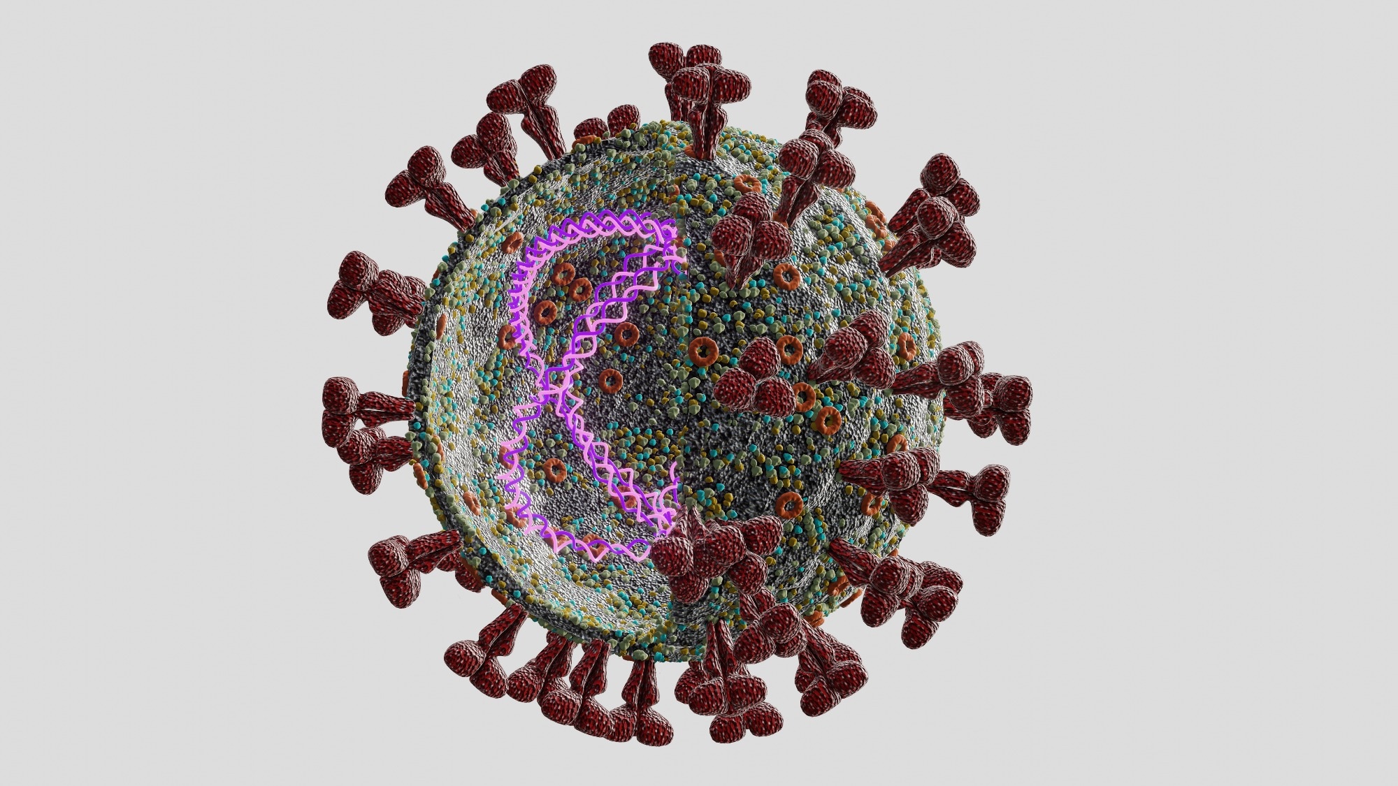
In a recent study posted to the bioRxiv* preprint server, researchers visualized the mechanism of action and inhibition of severe acute respiratory syndrome coronavirus 2 (SARS-CoV-2) non-structural protein 13 (NSP13) at high spatio-temporal resolution.

Background
Of all 15 SARS-COV-2 NSPs, NSP13, a ribonucleic acid (RNA) helicase, is critical for its replication. However, there are no presently approved antivirals that target nsp13. Unlike SARS-CoV-2 structural proteins, the amino acid sequence of nsp13 is one of the most conserved among many coronavirus (CoV) species (e.g., Middle Eastern respiratory syndrome CoV) and SARS-CoV-2 variants of concern (VOCs), including Omicron. Together, this makes nsp13 an attractive broad-spectrum antiviral target with the potential to combat future CoV outbreaks.
Structure and biochemical studies have proposed that nsp13 is a superfamily 1B (SF1B) RNA helicase. It uses an inchworm mechanism for translocation along single-stranded (ss) nucleic acid (NA) substrates by which nsp13 likely unwinds NA duplexes. Given its small step size, single molecule techniques have not been able to decipher the speed at which nsp13 walks along its NA substrate. Such resolution could shed light on how inhibitory molecules affect their workings.
About the study
In the present study, researchers developed single-molecule picometer-resolution nanopore tweezers (SPRNT) to measure the steps of SARS-CoV-2 nsp13 motion on DNA strands. Additionally, they demonstrated how SPRNT could be used to determine the mechanism of action of a helicase inhibitor. The team designed a single Mycobacterium smegmatis porin A (MspA) nanopore within a phospholipid bilayer. A voltage applied across this membrane caused an ion current to flow through the nanopore, which attracted negatively charged NA through the pore.
Different NA bases within the nanopore caused unique ion current blockages, which could be decoded into the NA sequence. A helicase bound to the captured NA strand would come to rest at the pore-rim and pull the NA resulting in sequential ion current steps. The team resolved these into single-nucleotide steps on sub-millisecond time scales to observe helicase motion along NA. Simultaneously, they determined the NA sequence of the substrate in the helicase.
It is also noteworthy that SPRNT applied a force proportional to the applied voltage on the enzyme/NA complex, which assisted or resisted the nsp13 motion depending on the end of nanopore NA was tied. Furthermore, the team observed NSP13’s motion along NAs in the presence of the adenosine triphosphatase (ATPase) inhibitor ATPγS.
Study findings
The researchers recorded 2413 individual NSP13 translocation and unwinding events and 27,641 helicase steps. The study results confirmed that NSP13 translocated at ~1000 nucleotides per second along ssDNA and unwinded DNA duplexes at a speed of ~100 base pairs per second. The NSP13 translocation rate was ATP dependent, with the maximum reaction rate (Vmax) ranging from 600 to 3000 s−1 and Michaelis constant (Km) ranging from 100 to 700 µM for ATP, depending on the underlying sequence context within NSP13. Such wide variations in translocation rates at different DNA positions indicated that the NA base identity affected NSP13 translocation kinetics.
The study results also showed that the NSP13-DNA complex was less stable and more easily dislodged by the application of force. Varying the assisting force from ~24 picoNewton (pN) to ~44 pN at saturating ATP caused no marked change in the average translocation speed of NSP13. Further, it suggested that the NSP13 translocation was predominantly ATP-hydrolysis-driven motion.
The authors also noted that dsDNA duplex unwinding steps (on average) were nearly eight-fold longer than that of ssDNA translocation. Moreover, dsDNA unwinding was slower than ssDNA translocation, although their dwell times were correlated. A similar effect was observed in another study examining SF1A helicase PcrA using SPRNT. Intriguingly, SARS-CoV-2 RNA-dependent RNA polymerase (RdRp) and NSP13 forms a complex at ∼170 nt/s at 37 °C, similar to what was observed as NSP13 unwinding speed using SPRNT.
Further, the authors noted that ATPγS interfered with NSP13’s action via several different kinetic processes. The dominant mechanism, however, depended on the application of assisting force. Although ATPγS is not a viable drug candidate for NSP13, it demonstrated the power of SPRNT in examining the mechanisms of helicase inhibition. It identified three methods of NSP13 inhibition:
i) reducing its processivity,
ii) preventing its domain 1A and 2A from joining following nucleotide binding, and
iii) slowing the ATPγS hydrolysis more relative to ATP.
Conclusions
Overall, the study highlighted SPRNT as a valuable and powerful tool for examining the role of NSP13 within the SARS-CoV-2 replication and transcription complex (RTC). The SPRNT method also showed a superior ability to facilitate studying the kinetics of NSP13 translocation or any helicase, even without a duplex. Furthermore, SPRNT experiments could facilitate studying NSP13 on native SARS-CoV-2 sequences to shed light on specific sequence elements of the highly structured SARS-CoV-2 genome and their role in NSP13 regulation.
*Important notice
bioRxiv publishes preliminary scientific reports that are not peer-reviewed and, therefore, should not be regarded as conclusive, guide clinical practice/health-related behavior, or treated as established information.
- Sinduja K. Marx, Keith J. Mickolajczyk, Jonathan M. Craig, Christopher A. Thomas, Akira M. Pfeffer, Sarah J. Abell, Jessica D. Carrasco, Michaela C. Franzi, Jesse R. Huang, Hwanhee C. Kim, Henry D. Brinkerhoff, Tarun M. Kapoor, Jens H. Gundlach, Andrew H. Laszlo. (2022). Inhibition of the SARS-CoV-2 helicase at single-nucleotide resolution. bioRxiv. doi: https://doi.org/10.1101/2022.10.07.511351 https://www.biorxiv.org/content/10.1101/2022.10.07.511351v1
Posted in: Medical Science News | Medical Research News | Disease/Infection News
Tags: Adenosine, Amino Acid, Bases, Coronavirus, Coronavirus Disease COVID-19, DNA, Enzyme, Genome, Helicase, Ion, Membrane, Molecule, Nucleic Acid, Nucleotide, Nucleotides, Omicron, Polymerase, Protein, Respiratory, Ribonucleic Acid, RNA, SARS, SARS-CoV-2, Severe Acute Respiratory, Severe Acute Respiratory Syndrome, Structural Protein, Syndrome, Transcription

Written by
Neha Mathur
Neha is a digital marketing professional based in Gurugram, India. She has a Master’s degree from the University of Rajasthan with a specialization in Biotechnology in 2008. She has experience in pre-clinical research as part of her research project in The Department of Toxicology at the prestigious Central Drug Research Institute (CDRI), Lucknow, India. She also holds a certification in C++ programming.
Source: Read Full Article


