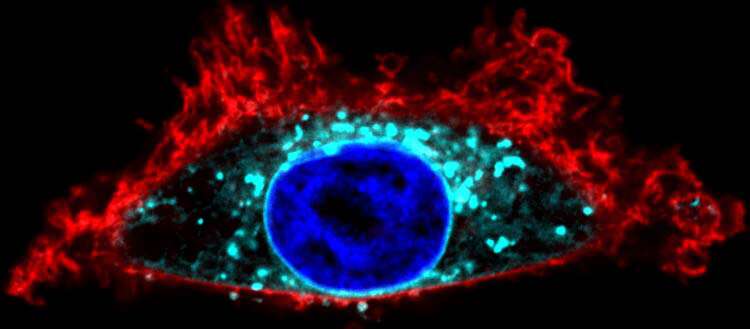
Peripheral membrane proteins temporarily bind to cell membranes, a necessary step for them to be able to fulfill their biological function. To do this, certain enzymes that catalyze their lipid modification come into action. This process is called palmitoylation. Some of these proteins can mutate and become oncogenic, such as the RAS proteins, which are responsible for about one third of cancers.
In order to understand how palmitoylation works and to use it for therapeutic purposes, scientists from the University of Geneva (UNIGE), Switzerland, developed a novel tool to visualize this process in living cells. They have shown that, contrary to what was thought, palmitoylation is possible not only in the Golgi apparatus, the usual site for protein processing and delivery, but also at the very site where the protein must act, e.g. the plasma membrane. This discovery, published in the journal Nature Communications, paves the way to innovative drug discovery strategies to target very precisely the membrane binding—and therefore activity—of oncogenic proteins.
Palmitoylation consist in the introduction of fatty acids into certain proteins for them to be able to bind to cell membranes. This mechanism is governed by precise rules, which depend on the sequence of each type of protein, and on the presence of specialized enzymes. Until now, the scientific community believed that palmitoylation of peripheral membrane proteins could only take place in one place in the cell, the Golgi apparatus.
“Indeed, these proteins are produced in the cytosol—the cell fluid—and then ‘swim’ to the Golgi apparatus, where they are modified before being transported to where they need to act,” explains Gonzalo Solis, researcher in the Department of Cell Physiology and Metabolism at the UNIGE’s Faculty of Medicine, and lead author of this work. “Nevertheless, we hypothesized the possibility of local palmitoylation, without passing through the Golgi apparatus. If this is true, it opens up completely new possibilities for the intervention of this mechanism.”
Observing and manipulating proteins
To test this hypothesis, the research team led by Vladimir Katanaev, professor in the Department of Cell Physiology and Metabolism and at the Center for Translational Research in Onco-Hematology at the UNIGE Faculty of Medicine, focused on a protein called Gαo, which is normally located at the plasma membrane and the Golgi apparatus.
The methodology used is completely new: “We brought the palmitoylating enzymes to a totally different compartment in the cell, the nuclear membrane,” explains Gonzalo Solis. “Gαo was recruited at the nuclear membrane, allowing us to identify the specific enzyme that palmitoylates them. We thus confirmed that this process can take place on the very site the protein is needed.”
Contrary to usual biochemical methodologies, the tool developed by Vladimir Katanaev and his team, which they named SwissKASH, is also the first to keep the cell alive and allows for the observation of the process dynamically. “Until now, there was no alternative to destroying the cell,” says Vladimir Katanaev. “Our method also makes it possible to determine exactly which protein reacts to which enzyme locally, which is essential if we want to control this mechanism for therapeutic purposes.”
A new drug target
Several peripheral membrane proteins, and in particular Gα subunit proteins and RAS proteins, are susceptible to mutation and thus acquire an aggressive oncogenic potential. Their oncogenicity depends on their ability to bind to the plasma membrane; palmitoylation thus plays a key role in the transformation of a healthy cell into a cancerous one.
Source: Read Full Article


