Exercise leaves muscles riddled with microscopic tears, so after a rigorous workout, the control centers of muscle cells — called nuclei — scoot toward these tiny injuries to help patch them up, scientists recently discovered.
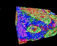
In the new study, published Oct. 14 in the journal Science, researchers uncovered a previously unknown repair mechanism that kicks in after a run on the treadmill. Striking images show how, shortly after the exercise concludes, nuclei scuttle toward tears in the muscle fibers and issue commands for new proteins to be built, in order to seal the wounds. That same process likely unfolds in your own cells in the hours after you return home from the gym.
The study authors discovered that “nuclei moved toward the injury site within 5 hours of injury,” Dr. Elizabeth McNally and Alexis Demonbreun, of the Northwestern University Feinberg School of Medicine, wrote in a commentary, also published in Science. And within only 24 hours of the injury, the repair process was “nearly complete.”
Skeletal muscles, which enable voluntary movements like walking, are made up of many thin, tubular cells; these cells are also called “muscle fibers,” due to their thread-like appearance. A single muscle can contain hundreds to thousands of muscle fibers, according to the National Cancer Institute. And each fiber contains units of contractile machinery, known as sarcomeres, that contract and lengthen during exercise.
Eccentric contraction, where your muscles are forcibly lengthened as they contract, can cause these sarcomeres to overstretch. (The second half of a bicep curl, where you slowly lower a dumbbell from shoulder-height to your side, and running downhill are examples of this type of exercise.) When sarcomeres overstretch during eccentric exercise, they can pull at the membrane surrounding them, causing damage, according to a 2001 review published in the Journal of Physiology.
In these situations, muscle cells rely on a skilled cellular pit crew to help fix them up. Previous studies have shown that, seconds after an exercise-induced injury occurs, various proteins form a “cap” over the damaged region of the membrane, and nearby mitochondria, the so-called powerhouses of the cell, help sop up any excess calcium that entered the cell through the tear, since the amount of calcium in muscle cells must be kept in check for them to function properly.
And now, the new study suggests that the nuclei in muscle cells rush over to help, too.
For the study, the researchers placed adult mice on a downward-tilted treadmill and then sampled muscle fibers from the animals following their jogging sessions. In addition, they asked 15 healthy human volunteers to run on a (person-sized) treadmill and then biopsied muscle fibers from the vastus lateralis, a part of the quadriceps.
They found that, in both the mouse muscle fibers and the human muscle fibers, proteins accumulated around tears in the fibers and formed “scars” within 5 hours after exercise. And in muscle fibers sampled 24 hours after exercise, clusters of nuclei had drawn close to the tears, whereas nuclei appeared farther away in the 5-hour samples. To see exactly how the nuclei had migrated toward the injury sites, the team grew mouse muscle cells in lab dishes and zapped them with lasers, to mimic exercise-induced injury.
In the lab-grown cells, the nuclei assembled around the laser injuries within 5 hours and soon generated “hotspots” of protein construction nearby. Specifically, the migration of nuclei was followed by a sudden explosion of mRNA molecules, a kind of genetic instruction manual built in the nucleus; mRNA essentially copies down the blueprints encoded in DNA and carries them out into the cell, where new proteins can be constructed. The newly built proteins then help to seal and reconstruct the injured muscle cells.
—Winter warriors: The fitness skills of 9 Olympic sports
—5 ways your cells deal with stress
—The twisted physics of 5 Olympic sports
In the future, medical treatments could potentially be devised to target the molecular pathways that allow the nuclei to migrate and start this repair process. That could help speed patients’ recovery after muscular injuries, McNally and Demonbreun wrote in their commentary.
Interestingly, the authors also found that mice that trained on the treadmill prior to the study developed fewer scars on their muscle fibers than mice that didn’t undergo any prior practice. This aligns with previous evidence that, with consistent training, muscles become stronger and less prone to tearing during trained movements, according to The New York Times.
Originally published on Live Science.
Nicoletta Lanese
Staff Writer
Nicoletta Lanese is a staff writer for Live Science covering health and medicine, along with an assortment of biology, animal, environment and climate stories. She holds degrees in neuroscience and dance from the University of Florida and a graduate certificate in science communication from the University of California, Santa Cruz. Her work has appeared in The Scientist Magazine, Science News, The San Jose Mercury News and Mongabay, among other outlets.
Source: Read Full Article


