Scientists use multicolor flow cytometry to identify and characterize targeted cellular subpopulations by using multiple fluorescent markers. The technology has seen rapid advancements in recent years leading to improvements in the method, resulting in a robust and reliable technique for performing complex cellular analysis rapidly and efficiently by evaluating several parameters simultaneously.
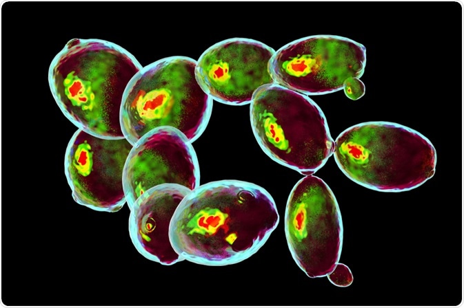
 Image Credit: Kateryna Kon / Shutterstock.com
In just seconds, the assay can investigate thousands of cells. Also, the repertoire of highly specific antibodies used for identifying substances is continually expanding. Further to this, more advanced flow cytometry instruments have been developed, allowing for the simultaneous analysis of over 20 parameters. As a result, multicolor flow cytometry has been instrumental in advancing the progress of research in the life sciences.
The basics of flow cytometry
Flow cytometry is the basic component of multicolor flow cytometry. In essence, the procedure has been designed to simultaneously analyze thousands of cells across multiple parameters, doing so in just a matter of seconds.
The process begins with placing a prepared sample into the cytometry instrument's flow chamber, where it enters a fluidics system. At this stage, the process of hydrodynamic focusing is used to separate the sample into its single-cell components. During this process, a controlled flow is introduced around the sample, forcing it into a narrow diameter and compelling the cells to separate.
Following this, the separated cells pass through a laser that records each individual cell as an event. On interaction with the laser, forward scatter (FSC) and side scatter (SSC) are produced by each cell, which is recorded by the instrument. In those cells that are fluorescently labeled, the fluorophore becomes excited as it passes through the laser and emits this light as it returns to its ground-state.
This emitted light is measured as it passes through a series of mirrors and filters until it reaches the appropriate detector for the targeted wavelength. These detectors, known as photomultiplier tubes (PMTs), recognize the fluorescence of a certain, predetermined wavelength.
In order to allow only the required light wavelengths to pass through, an optical filter (dichroic filter) is used to block out unwanted wavelengths and act as a mirror, which guides the specific wavelengths of light to the detectors.
Detection in flow cytometry
Flow cytometry relies on a combination of fluorophores and antibodies as detection reagents. Fluorophores can absorb and emit specific wavelengths of light from a wide range, depending on the fluorophore. This is known as the absorbance and emission spectra.
During flow cytometry, a specific wavelength of light is absorbed by the fluorophore. This light energy is transferred to the fluorophore's electrons, causing excitation. As the fluorophore returns to its ground-state, it releases the energy in the form of a photon of a longer wavelength than that which initially induced excitation.
The multicolor element of flow cytometry relies on multicolor panels that require the use of tandem dyes. Tandem dyes are made up of a donor fluorophore that generates excitation characteristics, and another acceptor fluorophore that creates the emission characteristics.
It is usual for the emission spectra of fluorophores to overlap, which can cause issues in reading the data because the overlap can generate a false-positive signal. Within multicolor experiments, this spectral overlap can be a particular problem, which means it must be controlled for by compensation. Compensation eliminates the chances of false positives from overlap by making sure that the detected fluorescence initiated from the measured fluorophore.
Methods of antigen detection
There are two methods of antigen detection in flow cytometry. The first is the direct method, where the primary antibody is directly conjugated to a fluorophore. The second is the indirect method, where the primary antibody is unconjugated and a fluorophore-conjugated secondary antibody directed against the primary antibody is used for detection.
There are benefits and drawbacks of each method; however, the direct method is most commonly used in multicolor experiments due to its comparative ease of production.
Multicolor experiment set-up
Building the panel is considered to be the most difficult part of the multicolor experiment set-up. First, the antigens that are of interest are selected. Then the lasers that have the corresponding wavelength to the fluorophores are selected.
Next, the cell population is considered. In cases where the antigen expression is unknown and/or low cell population is expected, a brighter fluorophore is used. Where a high antigen expression and/or high cell population is expected, a dimmer fluorophore is used.
After this, steps must be taken to minimize spectral overlap by sacrificing bright fluorochromes to avoid overlap and by using compensation to control for this.
Also, controls should be included in the set-up. Unstained cells can be used for determining negative populations, cell size, and granularity. Live/dead markers are useful in isolating healthy cells. Single-stained positive controls are used for setting compensation, and by considering the intensity of fluorescence minus one staining, positive populations can be identified.
Finally, the antibody concentration should be optimized by tiring the antibodies. This helps to avoid non-specific binding or reduced sensitivity.
Overall, multicolor flow cytometry has developed into a robust and reliable method for analyzing the subpopulations within a cell. As advancements in technology have been made, the method has improved, and researchers are benefiting from fast testing times and the ability to simultaneously analyze over 20 parameters at a time.
Sources:
- Baumgarth, N., and Roederer, M. (2000). A practical approach to multicolor flow cytometry for immunophenotyping. Journal of Immunological Methods, 243(1-2), pp.77-97. https://www.sciencedirect.com/science/article/pii/S0022175900002295
- Laskowski, T., Hazen, A., Collazo, R., and Haviland, D. (2019). Rigor and Reproducibility of Cytometry Practices for Immuno‐Oncology: A multifaceted challenge. Cytometry Part A. https://onlinelibrary.wiley.com/doi/10.1002/cyto.a.23882
- Maciorowski, Z., Chattopadhyay, P., and Jain, P. (2017). Basic Multicolor Flow Cytometry. Current Protocols in Immunology, 117(1). currentprotocols.onlinelibrary.wiley.com/doi/abs/10.1002/cpim.26
Further Reading
- All Flow Cytometry Content
- Flow Cytometry Methodology, Uses, and Data Analysis
- Flow Cytometry Techniques used in Medicine and Research
- Flow Cytometry History
- Fluorescence-Activated Cell Sorting
Last Updated: Dec 12, 2019

Written by
Sarah Moore
After studying Psychology and then Neuroscience, Sarah quickly found her enjoyment for researching and writing research papers; turning to a passion to connect ideas with people through writing.
Source: Read Full Article


