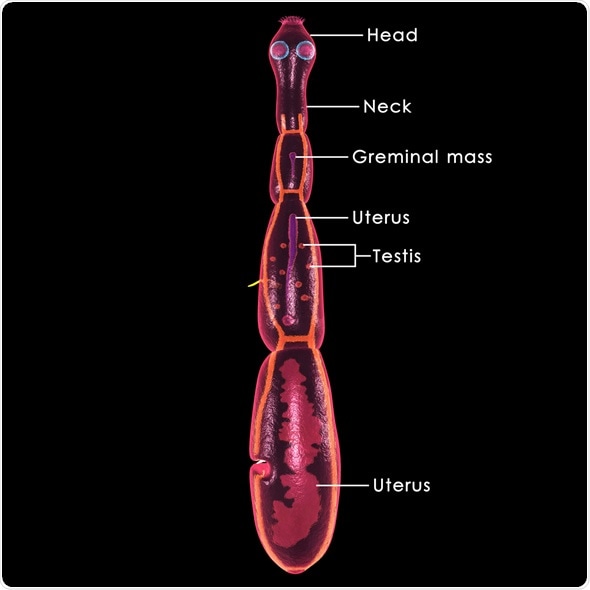
Cystic echinococcosis (Hydatid Disease) represents an emerging illness found in humans and livestock that arises as a result of eating infective eggs of the cestode parasite Echinococcus granulosus – a small tapeworm of carnivorous animals.
Also known as hydatid disease, this condition remains a significant cause of morbidity and mortality worldwide.
The ingestion of Echinococcus eggs results in the release of an early-stage larva (also known as the oncosphere) into the intestinal tract, with subsequent migration via blood or lymph to primary target organs such as liver or lungs.
At those locations, the oncosphere can mature into a vesicle which can grow by concentric enlargement, resulting in a fully mature hydatid cyst.
Cystic echinococcosis is not only responsible for severe disease and possible fatal outcomes, but also leads to high economic burden due to treatment costs, livestock losses and lost wages.
In the recent years noteworthy technological improvements have been made in characterizing, diagnosing, and treating infections with Echinococcus granulosus.
Clinical Presentation
Human cystic echinococcosis is characterized by a variable asymptomatic period during hydatid cyst growth, which depends on the patient age, site of cyst development, as well as the existence of a single or multiple cysts.
Approximately 70-80% of cysts are found in the liver, 10-20% in the lungs and only 5% in other sites in the human body (such as bones, spleen, central nervous system, or eyes).
Hydatid cysts can remain asymptomatic for life, especially if they stay small. If symptoms develop, they are most often associated with pressure effects of the cyst on surrounding tissues or organs. Non-specific signs that can appear include weight loss, anorexia, and weakness.
Nevertheless, traumatic rupture of the hydatid cyst is a serious manifestation that may lead to fatal anaphylaxis (i.e. a severe and extreme allergic reaction).
Other potential cyst complications include fistulas leading to bronchial or biliary obstruction, embolism of cyst content, and bacterial superinfections.
Diagnostic Approaches
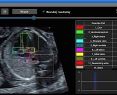
Due to its availability and convenience, the introduction of ultrasound in the clinical practice in the late 1970s has substantially improved the detection of cysts in the liver and abdomen.
World Health Organization developed a standardized classification system (pervasive in modern medicine) which divides hydatid cysts into six stages: CE1 and CE2 (active stage), CE3a and CE3b (transitional stage), and CE4 and CE5 (inactive stage).
Serology is also helpful, but its usefulness is limited by a lack of sensitivity and specificity.
Still, the combination of ultrasound technique and confirmatory serology represents a standard approach for epidemiological and clinical surveys. In unclear cases, diagnostic puncture of the cyst is performed by an experienced examiner.
Treatment and Prevention
In general, four different modalities are available in the treatment of hydatid disease: antihelminthic treatment, percutaneous drainage techniques, surgery, and “watch and wait” approach.
However, the management of patients depends not only on the individual case, but also on local expertise and available resources.
Surgery is indicated when there are large hepatic cysts (with multiple daughter cysts) or superficial hepatic cysts with the potential of spontaneous rupture.
Although antihelminthic treatment with benzoimidazole carbamates (i.e. mebendazole or albendazole) was once reserved for inoperable cases of human cystic echinococcosis, today they are more widely employed.
In this century, the percutaneous treatment known under the acronym PAIR (puncture, aspiration, injection, reaspiration) has secured an indispensable role in the treatment of hydatid disease.
In cases of small, non-complicated hepatic cysts, it is possible to consider the aforementioned “wait and see” approach as cysts can remain the same or stabilize even further.
The most effective preventive intervention against cystic echinococcosis is a combination of dog antihelminthic treatment and sheep vaccination.
Although successful control programs are important at the local level, the global distribution and public health significance of hydatid disease have not substantially changed.
Sources
- http://www.hindawi.com/journals/grp/2010/583297/
- http://www.hindawi.com/journals/ipid/2009/474368/
- http://www.who.int/mediacentre/factsheets/fs377/en/
- http://wwwnc.cdc.gov/eid/article/12/2/pdfs/05-0499.pdf
- http://www.ijidonline.com/article/S1201-9712(08)01440-9/fulltext
- http://www.thelancet.com/pdfs/journals/laninf/PIIS1473309907701342.pdf
- Gottstein B, Reichen J. Echinococcosis/Hydatidosis. In: Cook GC, Zumla AI. Manson’s Tropical Diseases, Twenty-Second Edition. Elsevier Health Sciences, 2009; pp. 1549-1568.
Further Reading
- All Cystic echinococcosis Content
Last Updated: Feb 26, 2019

Written by
Dr. Tomislav Meštrović
Dr. Tomislav Meštrović is a medical doctor (MD) with a Ph.D. in biomedical and health sciences, specialist in the field of clinical microbiology, and an Assistant Professor at Croatia's youngest university – University North. In addition to his interest in clinical, research and lecturing activities, his immense passion for medical writing and scientific communication goes back to his student days. He enjoys contributing back to the community. In his spare time, Tomislav is a movie buff and an avid traveler.
Source: Read Full Article