Stones found within the slender muscular tubes that carry urine from the kidneys to the bladder are referred to as ureteric calculi. These stones may be caused by factors such as a faulty diet, chronic urinary tract infections (UTIs) with or without abnormal urinary tract anatomy, metabolic dysfunction and hormonal imbalances.
 Credit: Suttha Burawonk/ Shutterstock.com
An inadequate fluid intake is perhaps the most important environmental factor in the etiopathogenesis of stone formation. Most stones are predominantly composed of calcium, but a good percentage is made of struvite (i.e. magnesium ammonium phosphate). Two other major categories of stones, which are less prevalent, are uric acid and cysteine stones.
The male-to-female ratio for the prevalence of urinary stones is 3:1. This distribution, however, is equal in children and in those whose stones arise due to hormonal or metabolic derangements. In contrast to these cases, stones that arise due to infection (struvite) are more likely to be found in women and are due to gram-negative bacteria that are capable of splitting urea into ammonia.
Likewise, women are more prone to developing serious renal complications such as hydronephrosis, which is swelling of the kidney that occurs due to urinary obstruction. Patients may present with nausea, vomiting, hematuria (i.e. blood in the urine) and pain that radiates to the back, flank and/ or lower abdomen, depending on where the stone is lodged within the ureters. These signs and symptoms warrant an in-depth medical workup.
This is especially important, because ureteric calculi may mimic other medical conditions of the abdomen, such as appendicitis or abdominal aortic aneurysm, which are both medical emergencies. Patient sex, race, duration of pain as well as associated nausea and hematuria are clinical parameters taken into account to predict the likelihood of a stone.
Diagnosis
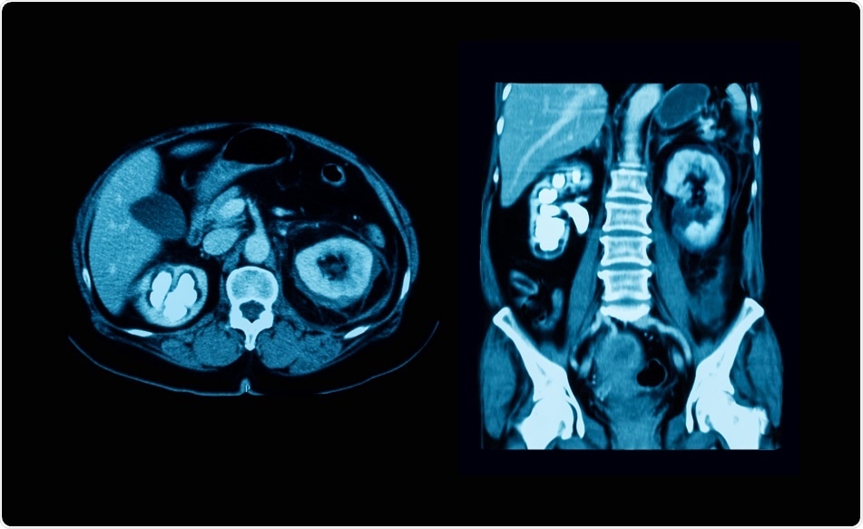
Although a computed tomography (CT) scan of the abdominopelvic region may be the imaging modality of choice for some, there are cases where ultrasound sonography of the urinary tract or radiographic images with or without contrast may be preferable. In addition to imaging tests, urinalysis and blood work may be ordered.
Urinalysis including leukocyte number, pH and bacteriuria is imperative for identifying the presence/ absence of hematuria and/ or infection. pH is particularly important, because increased urinary pH is suggestive of struvite, while a decreased pH increases the probability of finding uric acid stones. Blood work is also important to rule out systemic infection and more serious renal pathology.
Radiograph and CT
Plain urological radiography of the abdomen is also referred to as a kidney-ureter-bladder (KUB) radiograph or a flat plate. This imaging modality yields accurate results which help to locate the stone as well as its shape, size and composition. With regard to the latter, cysteine and uric acid calculi are radiolucent, whereas calcium stones are radiopaque. A further advantage of the KUB radiograph is that it is relatively quick to perform and inexpensive.
In contrast to the KUB radiograph, a CT scan is more expensive and exposes the patient to much higher doses of radiation. Moreover, smaller stones are difficult to see with CT scans and these scans may not identify some radiolucent calculi. To escape these disadvantages, the CT scan may be combined with KUB radiograph, based on many recommendations.
Nonetheless, the CT scan is an excellent imaging modality to help rule out more sinister pathologies, like abdominal aortic aneurysm, and, like the KUB radiograph, it can be used to determine the properties of the stone. This latter advantage is crucial for planning therapy.
Ultrasound and other diagnostic tests
Ultrasound is another imaging modality used to assist with diagnosing ureteric stones. It is especially employed in pediatric patients and in pregnant women, because modalities like the CT scan, which expose the patient to high doses of radiation, are contraindicated in the latter population. Ultrasound is also useful when there are suspected renal complications like hydronephrosis.
Other diagnostic tests that can be used in the diagnosis of ureteric calculi are intravenous pyelography and renal nuclear scan (i.e. use of radioisotopes). Pyelography with the help of a contrast agent can help outline the urinary system, but it comes at the cost of a higher risk of potential allergic reactions and adverse renal effects.
Sources:
- uroweb.org/wp-content/uploads/EAU-Guidelines-Urolithiasis-2015-v2.pdf
- http://pmj.bmj.com/content/postgradmedj/27/305/128.full.pdf
- https://www.ncbi.nlm.nih.gov/pmc/articles/PMC2600100/
- https://radiopaedia.org/articles/ureter
- https://www.ncbi.nlm.nih.gov/pmc/articles/PMC2518455/
Further Reading
- All Urinary Tract Infection Content
- What is a Urinary Tract Infection?
- Urinary Tract Infection Symptoms
- Urinary Tract Infection Diagnosis
- Urinary Tract Infection Treatment
Last Updated: Feb 27, 2019

Written by
Dr. Damien Jonas Wilson
Dr. Damien Jonas Wilson is a medical doctor from St. Martin in the Carribean. He was awarded his Medical Degree (MD) from the University of Zagreb Teaching Hospital. His training in general medicine and surgery compliments his degree in biomolecular engineering (BASc.Eng.) from Utrecht, the Netherlands. During this degree, he completed a dissertation in the field of oncology at the Harvard Medical School/ Massachusetts General Hospital. Dr. Wilson currently works in the UK as a medical practitioner.
Source: Read Full Article