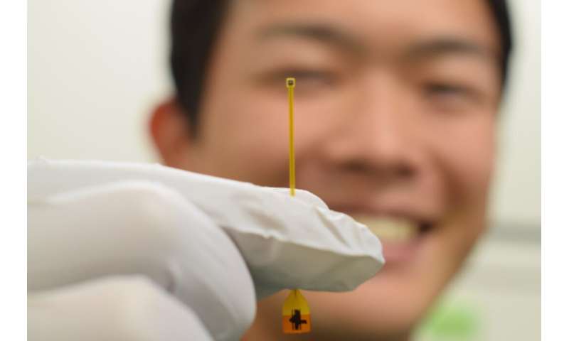
is stacked on an amplifier module (backside block) via a flexible interposer. Credit: Toyohashi University of Technology.")
A research team at the Department of Electrical and Electronic Information Engineering, Department of Computer Science and Engineering, Department of Applied Chemistry and Life Science, and the Electronics-Inspired Interdisciplinary Research Institute (EIIRIS) at Toyohashi University of Technology, and the National Institute of Technology, Ibaraki College has developed a < 3-μm-diameter ultra-small needle electrode with an amplifier for extracellular recordings. The team proposed an assembly technique via a single needle-electrode-topped amplifier package called STACK within a device geometry of approximately 1 × 1 mm2.
The STACK device enabled the recording of neuronal activity from a mouse’s brain in vivo with a high signal-to-noise ratio (SNR). Because of the significant advantage of the small needle geometry compared to conventional electrodes, the STACK device offers high biocompatibility and minimized tissue damage during recording, as well as further long-term and safe chronic recordings, toward the next generation of electrode technology in electrophysiology.
Microneedle electrode devices have been used as a powerful means of understanding how the brain works. However, the needle’s geometry should be further miniaturized in terms of biocompatibility and chronic application to avoid tissue damage: (i) Geometry of approximately 50 μm enhances the blood-brain barrier breach; (ii) > 20 μm causes a distribution of the local communication between glia; and (iii) < 10 μm produces no major traumatic tissue injury. Although advantages in micro/nano-scale fabrication technologies enable the formation of an approximately 10 μm needle-electrode, a smaller needle electrode causes electrical property degradation, making it impossible to record neuronal signals.
The research team has overcome these limitations by using an assembly technique by which a module with a <3-μm-diameter needle electrode is stacked on an amplifier module, called the single needle-topped amplifier package (STACK) device.
“To improve the electrical properties of our microscale silicon-needle electrode with a less than 10 μm diameter, we had proposed additional deposition process of low-impedance material to the needle’s tip, and demonstrated the neuronal recordings from rats with a 7-μm-diameter needle and from mice with a 5-μm-diameter needle. However, the deposition process enhances needle size, limiting the needle’s miniaturization. To tackle this challenge, we used a different approach, in which a small amplifier was embedded at the end of a small needle electrode. We confirmed the amplification effect in the neuronal recording, as neuronal signals were detected with the amplifier, but no signal was detected without. This result leads us to further needle miniaturization,” explains the first author of the article, master student Yuto Kita.
The leader of the research team, Associate Professor Takeshi Kawano, said, “Before this device concept, we first tried another fabrication method in which the silicon-needle electrode was integrated with the amplifier on the same silicon substrate. However, the fabrication technology did not work well because of the process mismatch between the silicon needle fabricated with high-temperature silicon growth (vapor-liquid-solid growth) on the (1×1)-silicon substrate and the MOSFET on (100)-silicon. The device package technology reported in this manuscript first appeared in our weekly group meeting, where I discussed it with students and postdoc members. Immediately after, we named the device technology STACK, which became our project code to pursue it.
Source: Read Full Article