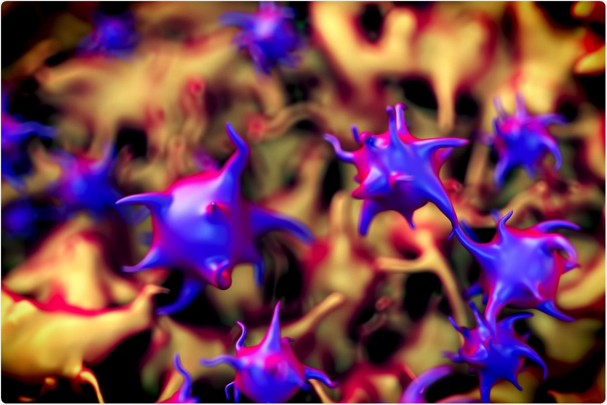
A major factor in coronavirus disease 2019 (COVID-19)-associated mortality is the development of coagulation abnormalities that may result in thromboembolism. Human platelets have been found by previous studies to respond to COVID-19 in a proinflammatory and procoagulatory manner, induced both directly by the virus or as a result of secondary interactions with antibodies and other activated biomolecules.
While this phenomenon is also observed in mouse models, the complete characterization of altered platelets has yet to be undertaken in mice, limiting the extrapolation of observations to humans. In a paper recently uploaded to the bioRxiv* preprint server, humanized ACE2 mice are infected with severe acute respiratory syndrome coronavirus 2 (SARS-CoV-2), and platelets are subsequently isolated from the blood for quantitative proteomic analysis.

Early platelet activation
Heterozygous humanized ACE2 (K18-hACE2) mice and C57BL/6J mice were inoculated with wildtype SARS-CoV-2 intranasally, with clinical assessments made daily for the following week. The group note that humanized mice experienced a greater loss of body weight and lower average body temperature one week post-infection compared to non-humanized mice, also experiencing several deaths by this time point.
Lung and kidney tissues were collected from the mice and viral loads determined at days two and four post-infection, with evidence of mononuclear cell infiltrates and low numbers of neutrophils in alveolar spaces. The SARS-CoV-2 spike protein was found within the alveolar epithelium, as well as CD61+ platelet aggregates in the lung and kidney capillaries. As these platelet aggregates are present amongst humanized mice as soon as two days post-infection, the group suggests that platelet activation and aggregation occur in these mice well before signs of clinical decline.
Changes in platelet population profile
Platelets were also collected from humanized mice infected with SARS-CoV-2 or not at two and four days post-infection, separated from blood by centrifugation and characterized by mass spectrometry.
Significant changes in platelet type and distribution were seen as a result of SARS-CoV-2 infection, with the specific character of a platelet population also differing notably between time points. Protein expression associated with platelet generation was also investigated by the group, finding that platelet complement and coagulation pathways were upregulated at two days post-infection, while at four days post-infection, intracellular infection pathways were upregulated.
P-selectin, platelet endothelial cell adhesion molecule-1 (PECAM–1), plasminogen, von Willebrand factor (vWF), and thrombin are proteins involved in the activation and aggregation of platelets, all of which are significantly upregulated in infected humanized mice at two days post-infection. However, the group also notes an increase in anti-coagulation proteins tissue factor pathway inhibitor (TFPI), protein C, protein S1, and thrombomodulin (THBD), suggesting that both pro- and anti-inflammatory factors are dysregulated by SARS-CoV-2 infection. Histidine-rich glycoprotein plasma proteins were also upregulated significantly at both time points in response to viral infection. However, the group suggests that this protein may have a negative influence on platelet aggregation, encouraging adherence to the inflamed endothelium via cell surface heparan sulfate, then subsequently blocking anti-inflammatory factors from bonding.
SARS-CoV-2 alters platelet factor expression
Proteins typically upregulated in response to viral infection were highly expressed by humanized mice four days post-infection, corresponding to activation of the NOD-like, Toll-like, and RIG-I-like receptor signaling pathways and resulting in the expression of interferon-induced proteins. Platelet factor 4 was the most abundant platelet chemokine amongst infected mice, which is known to be released from α-granules of activated platelets in COVID-19 patients.
Chemokines such as C-X-C motif chemokine ligand 12 and 5, along with C-C motif chemokine ligand 8 were also upregulated at both time points, indicating that monocyte and macrophage recruitment at sites of infection was underway, potentially promoting the generation of platelet-leukocyte aggregates.
The STRING protein database was utilized to determine the protein-protein interaction network developing within the mice throughout the course of SARS-CoV-2 infection, with the authors noting high interdependence between factors, particularly between those evident at two days post-infection. vWF, thrombin, and platelet factor 4 proteins are proposed to act as the center of dysregulation, interacting with proteins that determine platelet activation and aggregation and thereby enhancing virus-induced cytopathology.
Neither SARS-CoV-2 viral proteins nor the ACE2 receptor were detected by mass spectrometry in platelet samples, which is generally reflected amongst human platelet samples where SARS-CoV-2 uptake appears sporadic, and independent of ACE2 uptake. This study has demonstrated that humanized ACE2 (K18-hACE2) mice create a suitable model in which to study the dynamics of platelet activation and aggregation during SARS-CoV-2 infection, observing that during the course of COVID-19 coagulation pathways dominate early, while interferon signaling comes to dominate later, acting to dysregulate normal platelet function.
*Important notice
bioRxiv publishes preliminary scientific reports that are not peer-reviewed and, therefore, should not be regarded as conclusive, guide clinical practice/health-related behavior, or treated as established information.
- Subramaniam et al. (August 20th, 2021). Platelet proteome analysis reveals an early hyperactive phenotype in SARS-CoV-2-infected humanized ACE2 mice. bioRxiv preprint server. doi: https://doi.org/10.1101/2021.08.19.457020, https://www.biorxiv.org/content/10.1101/2021.08.19.457020v1.
Posted in: Medical Science News | Medical Research News | Disease/Infection News
Tags: ACE2, Antibodies, Anti-Inflammatory, Blood, Capillaries, Cell, Cell Adhesion, Chemokine, Chemokines, Coronavirus, Coronavirus Disease COVID-19, Endothelial cell, Glycoprotein, Histidine, Intracellular, Kidney, Leukocyte, Ligand, Macrophage, Mass Spectrometry, Molecule, Monocyte, Mortality, Neutrophils, Platelet, Platelets, Protein, Protein C, Protein Expression, Receptor, Respiratory, SARS, SARS-CoV-2, Severe Acute Respiratory, Severe Acute Respiratory Syndrome, Spectrometry, Spike Protein, Syndrome, Thromboembolism, Virus

Written by
Michael Greenwood
Michael graduated from Manchester Metropolitan University with a B.Sc. in Chemistry in 2014, where he majored in organic, inorganic, physical and analytical chemistry. He is currently completing a Ph.D. on the design and production of gold nanoparticles able to act as multimodal anticancer agents, being both drug delivery platforms and radiation dose enhancers.
Source: Read Full Article


