Transmission electron microscopy (TEM) is a technique used to observe the features of very small specimens. The technology uses an accelerated beam of electrons, which passes through a very thin specimen to enable a scientist the observe features such as structure and morphology.
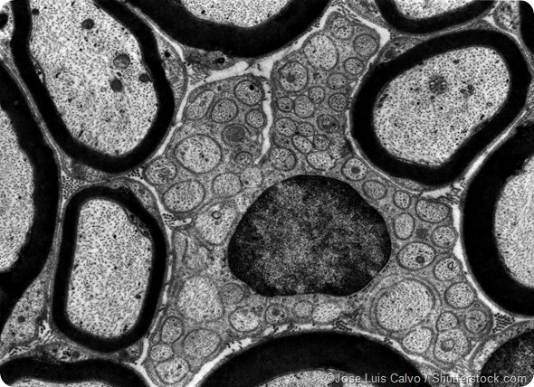
Transmission electron microscope (TEM) micrograph showing several peripheral myelinated fibers and a Schwann cell
The technology was first developed by German scientists Max Knoll and Ernst Ruska in 1931 and has evolved over the years to become a common technique that is used globally in science and engineering to look at micro and nanoparticles.
TEM can be used to observe particles at a much higher magnification and resolution than can be achieved with a light microscope because wavelength of an electron is much shorter than that of a photon. It also provides higher resolution images than a scanning electron microscope, which can only be used to scan and view the surface of a sample. Using TEM, scientists can be used to view specimens to the atomic level, which is less than 1nm.
Why transmission electron microscopy is used
This technology can tell us about the structure, crystallization, morphology and stress of a substance whereas scanning electron microscopy can only provide information about the morphology of a specimen. However, TEM requires very thin specimens that are semi-transparent to electrons, which can mean sample preparation takes longer.
How a transmission electron microscope works
In a transmission electron microscope, an electron gun, fires a beam of electrons. The gun accelerates the electrons to extremely high speeds using electromagnetic coils and voltages of up to several million volts.
The electron beam is focused into a thin, small beam by a condenser lens, which has a high aperture that eliminates high angle electrons. Having reached their highest speed, the electrons zoom through the ultra-thin specimen and parts of the beam are transmitted depending on how transparent the sample is to electrons.
The objective lens focuses the portion of the beam that is emitted from the sample into an image. Another component of the TEM is the vacuum system, which is essential to ensure electrons do not collide with gas atoms.
A low vacuum is first achieved using a either a rotary pump or diaphragm pumps which enable a low enough pressure for the operation of a diffusion pump, which then achieves vacuum level that is high enough for operations. High voltage TEMS require particularly high vacuum levels and a third vacuum system may be used.
The image produced by the TEM, called a micrograph, is seen through projection onto a screen that is phosphorescent. When irradiated by the electron beam, this screen emits photons. A film camera positioned underneath the screen can be used to capture the image or digital capture may be achieved with a charge-coupled device (CCD) camera.
Applications of a transmission electron microscope
This technology can be used in various industries from medical research where it is employed to investigate viruses and bacteria, for example, to forensic science, gemology and materials science.
References
- Wikipedia on transmission electron microscopy: https://en.wikipedia.org/wiki/Transmission_electron_microscopy#Three-dimensional_imaging
- Matter on transmission electron microscopy
- They Differ on the differences between transmission emission microscopy and scanning electron microscopy: http://theydiffer.com/difference-between-scanning-and-transmission-electron-microscope/
- About on the History of the Microscope: http://inventors.about.com/od/mstartinventions/a/microscope_2.htm
Further Reading
- All Comet Assay Content
- What is the Comet Assay?
- Comet Assay Applications
- All Microscopy Content
- Advances in Fluorescence Microscopy
Last Updated: Jan 25, 2019

Written by
Deborah Fields
Deborah holds a B.Sc. degree in Chemistry from the University of Birmingham and a Postgraduate Diploma in Journalism qualification from Cardiff University. She enjoys writing about the latest innovations. Previously she has worked as an editor of scientific patent information, an education journalist and in communications for innovative healthcare, pharmaceutical and technology organisations. She also loves books and has run a book group for several years. Her enjoyment of fiction extends to writing her own stories for pleasure.
Source: Read Full Article