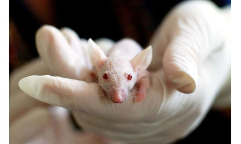
State-of-the-art photoacoustic microscopy (imaging using the heat given off by lasers) shows the blood flow in a mouse brain before, during, and after cardiac arrest, showing the drastic change in the brain’s blood flow when the heart stops and resurrects. Junjie Yao uses photoacoustic microscopy in his biomedical engineering research at Duke University.
This model could be a step toward visualizing human blood flow in new ways. In photoacoustic microscopy, a nanosecond laser pulse is used to excite molecules in the tissue and increase the local temperature by a very small amount. This heat causes the molecules at that site to expand and generate ultrasound emissions.
Scientists can then “listen for” the ultrasound signals outside the tissue using an ultrasound transducer and reconstruct where the photons are absorbed. Photoacoustic imaging can tremendously increase the penetration depth of traditional optical imaging, without any contrast agents. The resulting images are at a much higher resolution and at greater depths than those achieved by conventional microscopy. This makes photoacoustic imaging especially useful in biomedicine, given its precision and scalability.
https://youtube.com/watch?v=PO7I-r0ql_M%3Fcolor%3Dwhite
Source: Read Full Article


