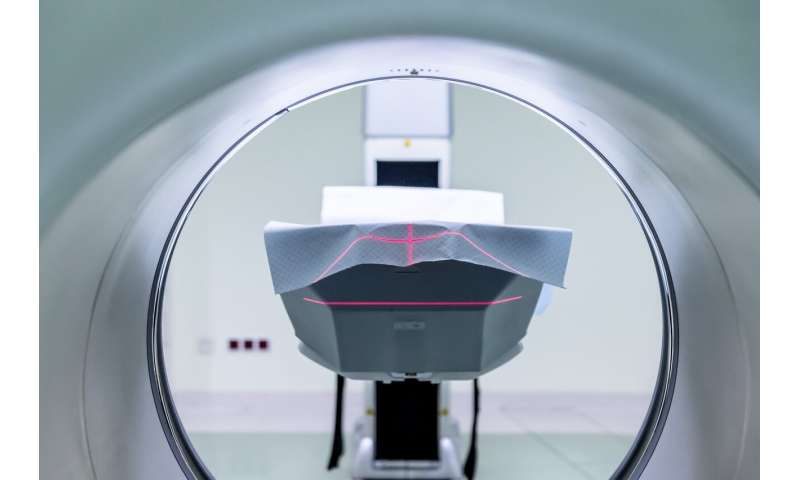
A new paper describes a breakthrough 100 micron resolution scan of the human brain that was created by a multidisciplinary team of Massachusetts General Hospital (MGH) researchers. The paper, which appeared in Scientific Data, highlights the highest resolution MRI scan of the whole human brain to be created to date, the images of which are 1,000 times more detailed than a standard clinical MRI scan.
“This unique dataset has a broad range of investigational, educational and clinical applications that will advance understanding of human brain anatomy in health and disease,” says Brian L. Edlow, MD, first author of the paper, associate director of the MGH Center for Neurotechnology and Neurorecovery (CNTR), and director of the Laboratory for NeuroImaging of Coma and Consciousness (NICC).
The MGH team included neuroscientists, neurologists, MRI physicists, engineers, computer scientists and anatomists. The project also required designing and building custom hardware to get the maximum possible resolution. MGH RF Coil Engineer Azma Mareyam designed the specialized MRI instrument for this project. “The RF coil used in a magnetic resonance scanner works as an antenna that transmits and receives signals from the patient or specimen,” she explains. “Those signals are then converted into images by other hardware/computer software.” The challenge in this case was to place many coils (32) close enough together that they could be fit snugly around the brain but would not interfere with each other. “This was done using many techniques we have developed in professor Lawrence Wald’s lab at MGH,” she explains.
The project has three main goals. First, it aims to generate insights into the structure of the brain, and connections between its parts. Another goal is to better understand the biology of neurological disorders such as traumatic brain injury and Alzheimer’s Disease through a new type of “network-based autopsy” that integrates MRI with histopathology. Finally, the team hopes to stimulate development of new, ultra-high resolution imaging techniques for use in living subjects.
The group, led by co-senior authors Bruce Fischl, Ph.D., and Andre van der Kouwe, Ph.D., has been working on this for over a decade, and in June 2019, released the 100 micron resolution MRI scan of a donated brain, including the underlying data, for which investigators already note promising additional uses.
In one application, for example, co-author Andreas Horn, MD, Ph.D., integrated the data from this scan into a software platform called Lead DBS, which may allow neurologists and neurosurgeons to improve the therapeutic outcome of deep brain stimulation electrode placement in a broad range of disorders, including Parkinson’s Disease and obsessive compulsive disorder. Another group, the Fiber Tractrography Lab at University of Pittsburgh, integrated the 100 micron MRI data with 3-dimensional tractography data, which show axonal pathways, to map the connectivity of the human brain. The data have also been integrated with a Neuroanatomy Atlas at Unochapecó University, Brazil.
There has been tremendous interest in this advanced application of MRI. Videos of the 100 micron MRI scan were viewed by more than 1 million people on Twitter, Youtube, Reddit, and Facebook within two weeks of release of the data in June.
Source: Read Full Article